
Blog, Physiotherapy, Posture
How to Read Your Medical Imaging Results (Without Freaking Out)
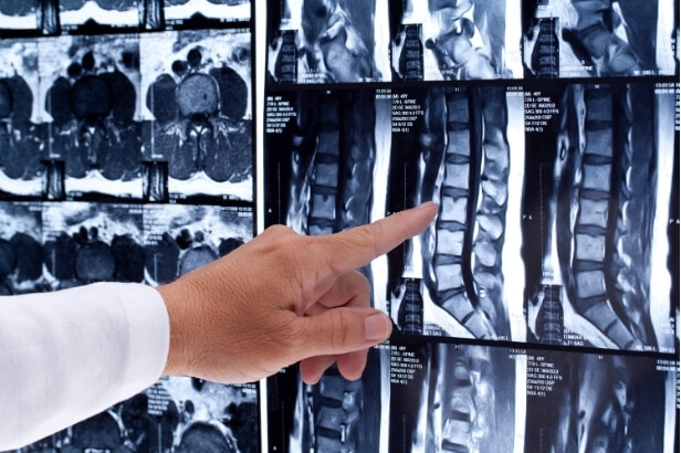
You’ve just received your medical imaging report. Now what?
It’s a scenario I see all the time: someone gets an MRI, CT, or X-ray for their pain, reads the report, and suddenly thinks their body is “broken.”
Words like “degeneration,” “disc bulge,” “osteophyte” or “narrowing” jump off the page and the worry sets in.
But here’s the truth: these findings are often completely normal and don’t necessarily explain your pain at all.
Why I’m Writing This
I’m Kevin Go, a physiotherapist with a strong interest in musculoskeletal pain, particularly neck, shoulder and lower back issues.
Many of my clients walk into the clinic with imaging results in hand, looking anxious and overwhelmed by the technical language.
This blog is here to help you understand your medical imaging, put things in context, and most importantly, avoid unnecessary fear.
Understanding the Stats & Situation
Here’s the reality that rarely gets explained to patients:
- A study in the American Journal of Neuroradiology found that over 50% of people in their 30s have disc bulges, even if they have no symptoms.
- By age 60, more than 80% of people will have some form of degenerative change in their spine and most of them feel fine.
- Many terms in scan reports sound serious, but they often reflect natural age-related changes, not damage or injury.
Your scan shows structure, but it doesn’t always explain pain.
The Problem
The major issue is that people read medical imaging reports without context. And when left to interpret it alone, it can lead to:
- Fear of movement or exercise
- Avoiding activities you actually need
- Stress, anxiety and even depression
- Belief that you’re “damaged” or need surgery, when you likely don’t
This cycle of fear can make your pain worse and your recovery slower.
What Can You Do Instead?
Here’s how to take control when reviewing your results:
Understand these common terms:
- Disc bulge/herniation – Often age-related and common in pain-free people
- Degenerative disc disease – Just like grey hair; part of ageing, not disease
- Osteophytes (bone spurs) – Bony changes that don’t always cause issues
- Narrowing/stenosis – Can be present without symptoms
- Tendinosis/tears – Many people have these with no pain at all
Ask yourself:
- Do the findings line up with your symptoms?
- Has a professional explained how these changes might (or might not) relate to your pain?
The Impact of Reframing
When you stop fearing your scan and start understanding it:
- You’re less likely to catastrophise
- You’ll move with more confidence
- You’ll focus on function, not just findings
- You’re more open to rehab and conservative treatment instead of rushing to surgery
In short, you’ll recover better.
Take Action Today
Here’s what to do if you’ve just received medical imaging results:
- Don’t Google it. The search results often lack context and can increase fear.
- Bring your report to a physio or GP who can interpret it properly.
- Focus on function. Your body’s ability to move and do what you love matters more than the image.
Key Takeaway
Your medical imaging is just one piece of the puzzle. Pain is complex, and imaging doesn’t always tell the full story. If you’ve been handed a report that scared you, take a breath. With the right guidance, the picture becomes much clearer.
Want to make sense of your scan results? Book in for an appointment, and let’s look at the whole picture together and not just what the scan shows.
 |
Written By:
Bachelor of Physiotherapy |
References
Greenberg, J.O. and Schnell, R.G. (1991) ‘Magnetic resonance imaging of the lumbar spine in asymptomatic adults’, Journal of Neuroimaging, 1(1), pp. 2–7. doi:10.1111/jon1991112.
Schmidt, C. (2017) ‘Systematic literature review of imaging features of spinal degeneration in asymptomatic populations’, manuelletherapie, 21(02), pp. 54–55. doi:10.1055/s-0043-105930.
Siivola, S.M. et al. (2002) ‘MRI changes of cervical spine in asymptomatic and symptomatic young adults’, European Spine Journal, 11(4), pp. 358–363. doi:10.1007/s00586-001-0370-x.
Tong, H.C. et al. (2006) ‘Magnetic resonance imaging of the lumbar spine in asymptomatic older adults’, Journal of Back and Musculoskeletal Rehabilitation, 19(2–3), pp. 67–72. doi:10.3233/bmr-2006-192-305.